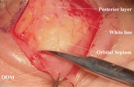
Fig. (3).
(Surgeon’s view) Detachment of the orbicularis oculi muscle from the posterior layer of the levator aponeurosis and the orbital septum. The white line is visualized. The orbital fat, which is seen through the translucent orbital septum, descents to the white line. OOM: orbicularis oculi muscle.

