
FIG. 3.
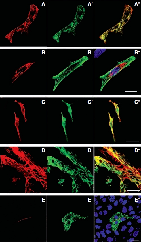
Detection of typical cardiac proteins in differentiated P19 cells by immunocytochemistry after 12 days of DMSO induction. (A–A″) P19 cell-derived cardiomyocytes that expressed cardiac troponin-T also expressed troponin-I. (A) Cardiac troponin-T. (A′) Troponin-I. (A″) Merged image. (B–B″) Representative images showing differentiated P19 cells expressing myosin heavy chain. (B) Myosin heavy chain. (B′) Troponin-I. (B″) Nuclear counterstaining with DAPI (blue) in the merged image. (C–C″) These cells also expressed MF20. (C) MF20. (C′) Troponin-I. (C″) Merged image. (D–D″) Tropomyosin (sarcomeric) expression in these same cells. (D) Tropomyosin (sarcomeric). (D′) Troponin-I. (D″) Merged image. (E–E″) Connexin43 was found among P19 cell-derived cardiomyocytes. (E) Connexin43. (E′) Troponin-I. (E″) Nuclear counterstaining with DAPI (blue) in the merged image. Scale bars: A, C, and D = 40 μm and B and E = 20 μm.