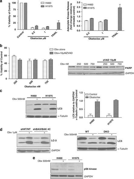
Figure 3.
Cell fate following obatoclax is determined by additional mechanisms other than apoptosis. (a) Obatoclax fails to induce loss in plasma membrane integrity. (left) H460 and H1975 NSCLC cells were treated at indicated doses of obatoclax for 72 h. H1975 cells were also treated with 20 ng/ml TNF-receptor-related apoptosis-inducing ligand (TRAIL). Viability of cells was assessed by ViaLight viability assay (Lonza Group Ltd.). Data are expressed as mean±S.D. (right) H460 and H1975 cells were treated as for ViaLight, however, this time toxicity of cells was assessed by ToxiLight cytotoxicity assay (Lonza Group Ltd.). Data are expressed as mean±S.D. (b) Inhibition of obatoclax-induced apoptosis by ZVAD.fmk fails to rescue cells. (left) H1975 cells were treated with 10 μM pan caspase inhibitor ZVAD.fmk for 30 min before treatment with indicated doses of obatoclax for 48 h. Viability of cells was assessed by ViaLight viability assay. Data are expressed as mean±S.D. (right). The effect of ZVAD on obatoclax-induced PARP cleavage was assessed by SDS–PAGE. GAPDH was used as a loading control. (c) Obatoclax induces significant processing of LC3. (left) H460 and H1975 cells were treated with 500 nM obatoclax for 48 h and the level of LC3 processing assessed by SDS–PAGE. (right) Quantification of the level of LC3 processing following obatoclax was achieved by densitometry analysis using ImageJ. Data are expressed as mean±S.D. (d) Obatoclax-induced LC3 processing is independent of BAX and BAK. (left) H460shBAX/BAK 4C cells were treated with 1 μM obatoclax for 48 h and the level of LC3 processing assessed by SDS–PAGE. GAPDH was used as a loading control. (right) Wild type and BAX/BAK DKO MEFs were treated with 500 nM obatoclax for 48 h and the level of LC3 processing assessed by SDS–PAGE. α-Tubulin served as a loading control. (e) Obatoclax induces de-phosphorylation of S6 kinase. H460 and H1975 cells were treated with 500 nM obatoclax for 48 h and the level of S6 kinase phosphorylation determined by SDS–PAGE and immunoblotting with a phospho-S6 kinase antibody that detects phosphorylation of thr389. GAPDH was used as a loading control

