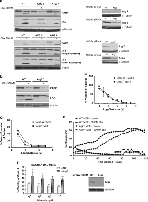
Figure 6.
Obatoclax-induced LC3 processing requires Atg7 and Atg5. (a) siRNA knockdown of Atg5/7 reduces obatoclax-induced LC3 processing. H460 (top) and H1975 (bottom) cells were transfected with 100 nM of Atg7 or Atg5 siRNA, or control non-targeting siRNA (NT) for 48 h, and then treated with 500 nM obatoclax for a further 48 h. The level of LC3 processing and PARP cleavage in Atg5/7-transfected cells relative to NT-transfected cells was then determined by SDS–PAGE. α-Tubulin and β-actin were used as loading controls. (b) Obatoclax-induced LC3 processing is lost in mouse embryonic fibroblasts derived from mice deficient for Atg7. Wild-type and Atg7−/− MEFs were treated with 500 nM obatoclax for 48 h and the level of LC3 processing and PARP cleavage was determined by SDS–PAGE. β-Actin was used as a loading control. (c) Autophagy is not essential for obatoclax-induced toxicity. The effect on the viability of Atg7−/− MEFs and their wild-type controls following obatoclax treatment for 48 h. EC50 values for each cell line were; Atg7 WT 84 nM; Atg7−/− 58 nM. Data are presented as mean±S.D. (d) Loss of Atg7 fails to rescue cells. Atg7 wild-type and Atg7−/− cells were plated in 24-well plates at single-cell densities and treated with indicated doses of obatoclax for 24 h. Media was replaced after 24 h and cells allowed to form colonies over 7–10 days to assess the effect of obatoclax on clonogenic growth. Data are presented mean±S.E. (e) Loss of Atg7 fails to rescue cells over the long term. Cells were seeded in 6-well plates and drugged at indicted doses. Cells were then monitored using the INCUCYTE live cell imaging system (Essen BioScience). Cell confluency was determined using calculations derived from phase-contrast images. This data was condensed using algorithms into quantified metrics to obtain kinetic proliferation curves. (f) siRNA knockdown of Atg7 rescues BAX/BAK DKO MEFs. DKO MEFs were transfected with 100 nM of Atg7 siRNA or control non-targeting siRNA (NT) for 48 h. Cells were then treated with indicated doses of obatoclax for a further 48 h and the effect on viability determined by ViaLight assay. Data are presented as mean±S.D. The level of knockdown was determined by SDS–PAGE. GAPDH was used as a loading control

