Figure 1.
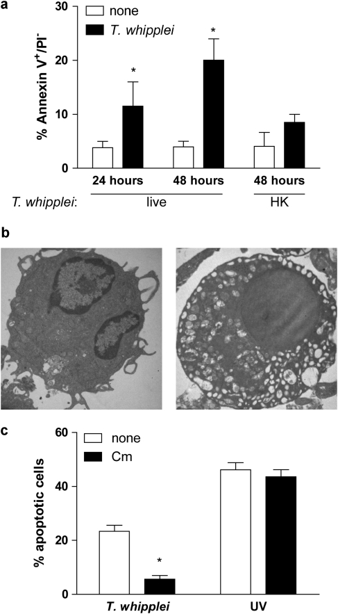
T. whipplei induces apoptosis of human MDM. (a) MDM were infected with live or heat-killed (HK) T. whipplei (MOI 50 : 1) for 4 h, washed and incubated for 24 and 48 h. Cells were then washed and stained with annexin V-FITC and PI and analyzed by flow cytometry. The data are the mean±S.D. of three independent experiments. (b) Uninfected (left) and T. whipplei-infected (right) MDM were observed by TEM after 48 h of incubation. (c) T. whipplei-infected MDM were incubated for 48 h in the presence or not of chloramphenicol (Cm, 20 μg/ml). As a control, MDM were exposed to UV light. Apoptosis was determined by TUNEL staining and quantified by examining 3–5 fields per condition (∼100 cells per field). The percentage of TUNEL-positive cells was calculated as the ratio between TUNEL-positive and DAPI-stained nuclei × 100. *P<0.05, Mann–Whitney's U-test

