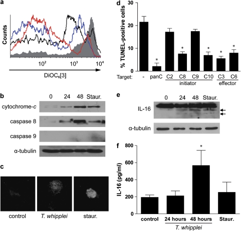
Figure 3.
T. whipplei triggers Δψm disturbance and caspase activation. MDM were infected with T. whipplei (MOI 50 : 1) for 4 h, washed and incubated for additional periods. As a control, MDM were treated with staurosporine (Staur) for 6 h. (a) Cells were incubated with DiOC6[3] and fluorescence was monitored by flow cytometry. The results are representative of two independent experiments (gray line: uninfected cells; black line: staurosporine-treated cells; blue and red line: MDM incubated 24 and 48 h after infection, respectively). (b) MDM lysates were analyzed by immunoblotting for cytochrome-c, caspase 8, caspase 9 or α-tubulin as loading control. One representative experiment is shown (n=2). (c) Caspase 3 activity was determined by immunofluorescence using active caspase 3-specific antibodies. (d) MDM were infected with T. whipplei (MOI 50 : 1) for 4 h, washed and incubated for 24 and 48 h in the presence of different caspase inhibitors. Apoptosis was determined by TUNEL staining and quantified by examining 3–5 fields per condition. The percentage of TUNEL-positive cells was calculated as the ratio between TUNEL-positive and DAPI-stained nuclei × 100. (e) MDM lysates were analyzed by immunoblotting for IL-16 and α-tubulin as loading control. Arrows represent the cleaved forms of IL-16. One representative experiment is shown (n=2). (f) MDM supernatants were assessed for IL-16 by ELISA. The data are the mean±S.D. of three experiments. *P<0.05, Mann–Whitney's U-test

