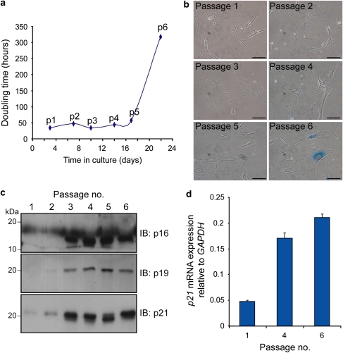
Figure 1.
MEFs undergo senescence after 5 passages. (a) At each passage (p), cells were counted and the doubling time was calculated. The p6 cells show greatly increased doubling time as cells enter senescence. (b) At each passage, the cells were analyzed for SA-β-gal staining. By passage 6, the majority of cells are blue, indicating that senescence has been activated. Scale bars=40 μm. (c) Equal amounts (50 μg) of MEF protein lysates loaded onto replicate gels were analyzed by immunoblotting. Typical markers of senescence p16, p19 and p21 are increased during passaging. (d) p21 mRNA expression in MEFs was quantitated by qPCR at passages 1, 4 and 6. Error bars show S.E.M. (n=3)

