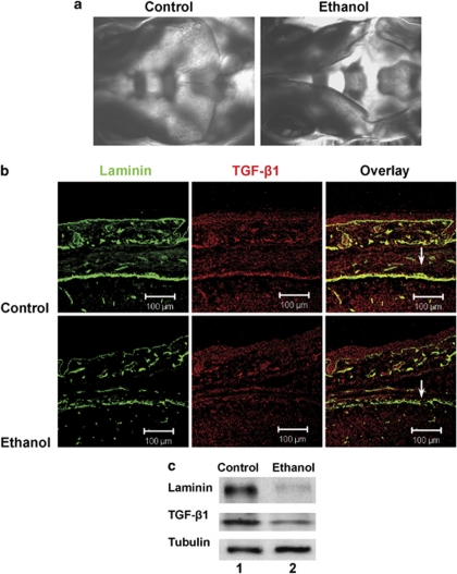
Figure 4.
Prenatal ethanol exposure leads to defects in parietal bone development and loss of laminin and TGF-β1 expression in the meninges. (a) Pregnant mice were intubated with 3 g/kg ethanol at days E8.5 and E9.5. At day E17.5, embryos were extracted and subjected to Alizarin Red staining (a) or coronal cryosectioning and immunocytochemistry for laminin (green) and TGF-β1 (red, b). Note ethanol-induced malformation of the parietal bone. Also note disrupted laminin staining and loss of TGF-β1 in ethanol-exposed embryos (arrows). (c) Immunoblot showing reduced levels of laminin and TGF-β1 in 20% of embryos, concurrent with defects in head development. This experiment was performed three times with a total of 25 embryos from isocaloric control and ethanol-intubated pregnant mice. Lane 1, embryo from control mouse, lane 2, embryo from ethanol-treated mouse

