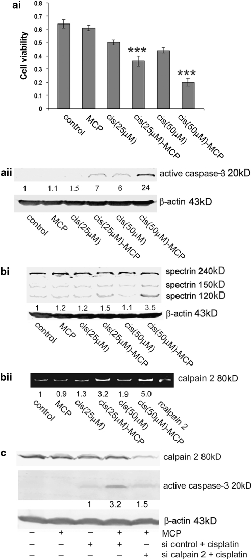
Figure 7.
The blockage of Gal-3 by GCS-100/MCP enhanced cisplatin-induced apoptosis in a calpain-dependent way. (ai) Cell viability assay by using MTT. Columns (n=3), mean; bars, ±S.D. ***P<0.001 compared with cisplatin treatment only. (aii) Western blot analysis of active caspase-3. Numbers represent the relative intensity of active caspase-3 normalized to β-actin. The value of control cells was set as “1”. Parental PC3 cells were pretreated with 0.3% MCP for 30 min and then cisplatin for 24 h. (bi) Calpain activity as represented by western blot analysis of spectrin 150 kD. Numbers represent the relative intensity of spectrin 150 kD normalized to β-actin. The value of control cells was set as “1”. (bii) Casein zymography to detect calpain activity. Purified recombinant rat calpain 2 was used as the positive control. Numbers represent the intensity of calpain 2. The value of control cells was set as “1”. Parental PC3 cells were pretreated with 0.3% MCP for 30 min and then cisplatin for 12 h. (c) Western blot analysis of active caspase-3 showed that calpain 2 knockdown attenuated the proapoptotic effect of Gal-3 blockage by GCS-100/MCP. Parental PC3 cells were transfected with siRNA duplex targeting calpain 2 or non-target control siRNA. After 24 h of transfection, cells were pretreated with 0.3% MCP for 30 min, and then treated with cisplatin (50 μM) for 24 h. Numbers represent the relative intensity of active caspase-3 normalized to β-actin. The value of cells transfected with control siRNA plus cisplatin treatment was set as “1”. Data are representative of three independent experiments

