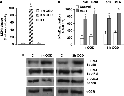
Figure 1.
(a) Primary cortical neurons were exposed to 1 h OGD and then 24 h later to 3 h OGD. The next day, cell viability was measured by LDH assay. Sub-lethal ischemic injury totally prevented the 3 h OGD-mediated neurotoxicity. Bars are means±S.E.M. of three separate experiments run in triplicate; *P<0.01 versus the corresponding control value. (b) Activation of p50 and RelA was evaluated by ELISA analysis in nuclear extracts from cortical cells exposed to 1 or 3 h OGD. Bars are means±S.E.M. of three separate experiments; *P<0.05 versus the corresponding control value. (c) Representative picture of co-immunoprecipitation analysis of p50, RelA and c-Rel dimers in nuclear extracts from primary cortical neurons showed a high level of p50/RelA complex activation after 1 or 3 h OGD, whereas the activation of c-Rel dimers decreased slightly

