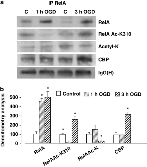
Figure 2.
(a) Immunoprecipitation analysis of RelA acetylation and association with CBP in nuclear extracts of primary cortical neurons exposed to 1 or 3 h OGD. RelA Lys310 acetylation decreased after 1 h OGD and increased after 3 h OGD. Total RelA acetylation was not altered by 1 h OGD but was reduced by 3 h OGD. RelA association with CBP increased in nuclear extracts of cells subjected to 3 h OGD. The signal given by IgG(H) was used as a control for the quality of the immunoprecipitation. Similar results were obtained in at least four separate experiments. (b) Values from densitometric analysis of immunoblot bands are expressed as a percentage of the corresponding control value. Error bars depict means±S.E.M.; *P<0.05 versus control

