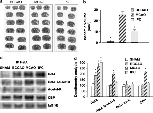
Figure 3.
(a) Representative images of infarct areas in brain coronal sections of mice exposed to BCCAO for 5 min or to MCAO for 20 min. In preconditioning experiments (IPC), mice were subjected to BCCAO and the next day to MCAO. Ischemic lesions were evaluated 3 days later. (b) Infarct volume in mice subjected to BCCAO, MCAO or IPC. Data are reported as means±S.E.M. (n=9 or 10 animals per group); *P<0.05 versus MCAO value. (c) Representative picture of co-immunoprecipitation analysis of RelA acetylation in nuclear extracts of mice exposed to BCCAO, MCAO or IPC (n=3 per group). Nuclear extracts were prepared 4 h after the end of each experimental condition. Acetylation on the Lys310 residue, as well as levels of the RelA/CBP complex, increased in mice exposed to MCAO. (d) Densitometric analysis of immunoblot bands. Values are expressed as a percentage of the control (Sham) value. Error bars depict means±S.E.M.; *P<0.05 versus control

