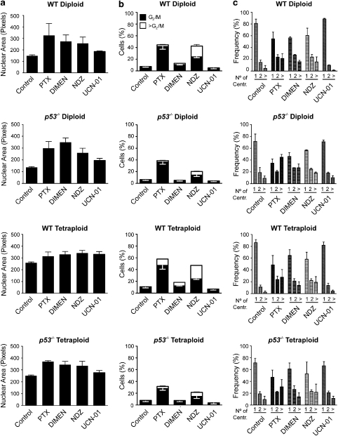
Figure 5.
Automatic analysis of fluorescence microphotographs allows for the assessment of polyploidization. Human colon carcinoma HCT 116 cells with the indicated ploidy and p53 status stably expressing fusion proteins for the detection of chromatin status (H2B-GFP) and centrosome number (DsRed-Centrin) were left untreated (Control) or incubated with paclitaxel (PTX), dimethylenastron (DIMEN), nocodazole (NDZ) or 7-hydroxystaurosporine (UCN-01) for 72 h. (a) Automated analysis of fluorescence microphotographs allowed for the quantification of nuclear area. Columns depict the mean of the median values±S.E.M., as recorded in three independent experiments. Mitotic blockers (but not UCN-01) provoked a relevant increase in the nuclear area of both diploid and tetraploid cells, irrespective of their p53 status. (b) Cytofluorometric analysis upon Hoechst 33342 staining confirmed that, in clear contrast to UCN-01, mitotic inhibitors (in particular PTX and NDZ) led to the accumulation of cells with a DNA content ⩾G2/M, in both diploid and tetraploid cells. As at this time point mitotic cells were not detectable by imaging (data not shown), these events represented bona fide aneuploid cells. (c) Automated quantification of the number of centrosomes per cell (No. of Centr.) suggested that p53−/− HCT 116 cells tended to accumulate supernumerary centrosomes more readily than their wild-type (WT) counterparts. In (b) and (c), data are mean values±S.E.M. (n=3 independent experiments)

