Figure 3.
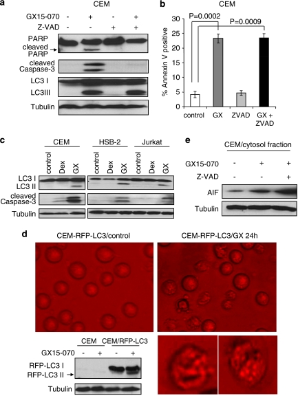
Apoptosis and autophagy are induced by GX15-070 treatment. (a) CEM cells were treated with 100 nM GX15-070±50 μM Z-VAD for 32 h. Z-VAD was added 30 min before GX15-070 treatment. Cleavage of PARP or caspase-3 and LC3 conversion were detected by western blot analyses. (b) CEM cells were treated with 100 nM GX15-070 in the presence or absence of 50 μM Z-VAD for 48 h. The percentage of cell death was determined by Annexin V-PI staining followed by FACS analysis. Values represent the mean±S.D. of three independent experiments. (c) Dex-sensitive CEM or -resistant HSB-2 and Jurkat cells were treated with 100 nM Dex or 100 nM GX15-070 for 24 h, and LC3 conversion and caspase-3 cleavage were detected by western blot analyses. (d) CEM cells were stably transfected with RFP-LC3, and G418-resistant clone cells were then treated with 100 nM GX15-070 for 24 h. Punctated RFP-LC3 was visualized under a fluorescent microscope. Western blot analysis confirmed the conversion of RFP-LC3. These pictures are representative of three independent experiments. Magnification is × 40. Bottom pictures are enlarged single-cell images. (e) CEM cells were treated with 100 nM GX15-070±50 μM Z-VAD for 24 h and release of cytochrome c or AIF in cytosol fraction was determined by western blot analyses

