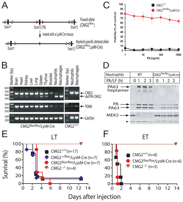
Figure 2. Generation and anthrax toxin sensitivity of the myeloid-specific CMG2-null mice.
(A) Schematic targeting strategy. Diagram of the CMG2flox allele having exon 12 flanked by LoxP sites (“Floxed”), and the myeloid-specific CMG2-null allele (CMG2flox/LysM-Cre). The homozygous myeloid-specific CMG2-null mice (CMG2flox/flox/LysM-Cre) were obtained by the intercrossing of CMG2+/flox/LysM-Cre mice. The red arrowheads indicate LoxP sites.
(B) RT-PCR analyses of the CMG2 TM domain deletion in various tissues, macrophages, and neutrophils in a CMG2flox/flox/LysM-Cre mouse. Macrophages and neutrophils from a WT mouse were also included as controls. See Figure S1 for neutrophil isolation.
(C) Toxin susceptibility of BMDMs from WT and myeloid-specific CMG2-null mice. BMDMs were treated with various concentrations of PA plus FP59 (100 ng/ml) for 48 h. Cell viability was evaluated by MTT assay. Data are reported as mean viability ± S.D.
(D) Binding and processing of PA on mouse neutrophils. Neutrophils isolated from mouse bone marrow were incubated with 1 μg/ml PA and 0.5 μg/ml LF at 37 oC for 0–3 h. Cells were washed, lysed, and cell lysates subjected to Western blotting using either an anti-PA antiserum or a MEK2 antibody. A non-specific cross-reactive band indicated by the arrow head at the lower left of the blot serves as protein loading control.
(E-F) Sensitivity of myeloid-specific CMG2-null mice to the anthrax toxins. CMG2flox/flox/LysM-Cre mice and their control mice were treated with 100 μg PA plus 100 μg LF (intraperitoneally) (E) or with 50 μg PA plus 50 μg EF (intravenously) (F) and survival monitored for 2 weeks. Whole body CMG2-null mice (CMG2−/−) in the same genetic background (C57BL/6) were included as additional controls.

