
Figure 5.
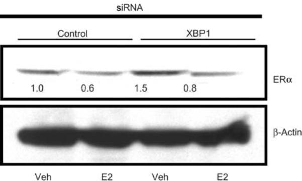
ERα levels in MCF7 cells treated with control or XBP1 siRNA. MCF7 cells were transfected with control or XBP1 siRNA and subsequently treated with vehicle or E2 for 24 h. ERa levels were assessed by Western blotting. Levels of β-actin are shown as loading control. The Western blots were scanned and quantified. Levels of ERα protein normalized for β-actin, relative to control siRNA-vehicle treated cells, are indicated below each band.