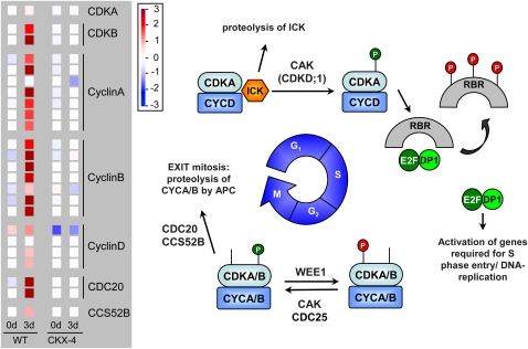
Figure 11.
Expression of cell cycle genes in wild-type (WT) and CKX-4 tubers after GA3-mediated tuber sprouting. Expression data of differentially expressed ESTs representing genes of the cell cycle are shown in the left-hand panel and a schematic overview of the mitotic cell cycle, modified after Inzé and De Veylder (2006), is shown on the right. Color bars as displayed by the MapMan tool were copied to and aligned in Microsoft PowerPoint. The color scale next to the panel indicates transcript levels, with red representing an increase and blue representing a decrease in transcript levels. The colors saturate at 8-fold change. Details on the assignment of individual POCI identifiers to genes shown are listed in Supplemental Table S3. At the G1/S-phase transition, CDK inhibitory protein ICK dissociates from CDKA-CYCD complexes and is degraded via the proteasome pathway. Phosphorylation of CDKA then activates the CDKA-CYCD complex, which initiates phosphorylation of the retinoblastoma-related protein (RBR), releasing the E2F-DP1 complex. This complex promotes the transcription of genes required for progression into and through S-phase, where DNA replication takes place. The G2/M-phase transition is accompanied by a marked increase in transcription of both A- and B-type CDKs and A- and B-type cyclins. CDKA/B-CYCA/B complexes are activated by removal of the inhibitory phosphate by CDC25 and phosphorylation by CDK-activating kinase (CAK), both enzymes acting on the CDK part of the complex. The reverse reaction, inhibitory phosphorylation of CDK, is catalyzed by kinase WEE1 and promotes endoreduplication. During mitosis, CDC20- and CCS25-activated anaphase-promoting complex (APC) degrades cyclins A and B, leading to the transition from mitosis into G1-phase. CDC, Cell division cycle; DP, docking protein.

