
Figure 4. Cx43-S368 phosphorylation on the actin bridges.
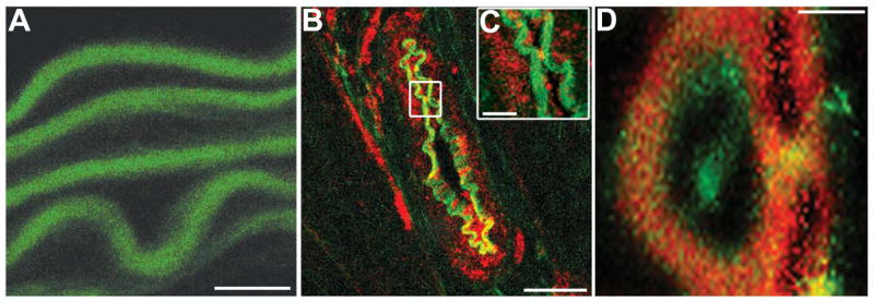
In A–C, green is IEL, red is Cx43-S368. A, there is no Cx43-S368 detectable in the aorta. However, in B–C, transverse sections of mouse cremasteric arterioles demonstrate heavy staining for Cx43-S368 in EC and VSMC. C, is an enlarged image from B (white box). D, actin bridges are highlighted by phalloidin (red); Cx43-S368 (green) is clearly seen between cell types. Bar in A is 40 μm, bar in B is 20 μm; bar in C is 10 μm; and bar in D is 5 μm.