Figure 1.
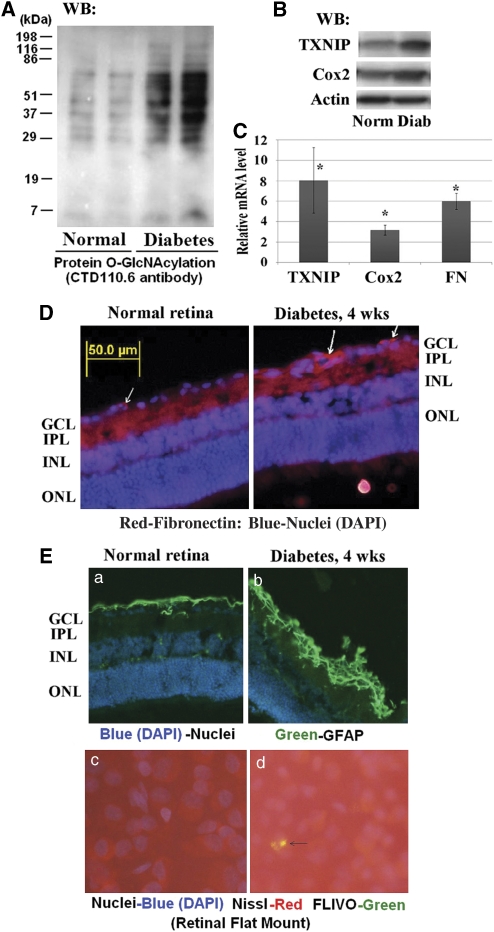
Diabetes increases HBP, TXNIP expression, inflammation and fibrosis in the retina. (A, B) Cellular protein extracts (30 μg) were analyzed for protein Ser or Thr-O-GlcNAcylation using CTD110.6 antibodies, which specifically recognize the O-GlcNAc moieties, and TXNIP expression on western blots. In addition, Cox-2 expression was also determined. (A) Diabetes induces HBP flux as observed by enhanced CTD110.6 reactivity in diabetic retina (representative blots from n=5). At present, we do not know the identity of these proteins. (B) Similarly, diabetes also increases TXNIP protein levels (∼3.6±0.5-folds, P<0.01 versus normal retina; n=6). Furthermore, Cox-2 level is also induced in the diabetic retina (∼1.67±0.17-folds, P<0.05 versus normal retina, n=5). Actin is not changed. (C) RT-qPCR analysis reveals that TXNIP expression is significantly increased in the diabetic retina (8.06±3.18-folds) over normal retina, which correlates with increases in Cox-2 (3.18±0.49) and FN (5.99±0.78) expressions. The asterisk symbol represents significant change (P<0.01, n=7) in the diabetic retina when compared with normal retina. (D) On immunohistology, FN expression is increased in the basal membrane of blood vessels in GCL and also in the ILM as shown by the arrows. A representative of n=3 is shown. (E) Diabetes induces glial reactivity and ganglion injury in early DR. Immunohistology analysis reveals that GFAP expression is increased in the diabetic retina (a, b), which correlates with caspase-3 activation in the diabetic retina as demonstrated by positive staining for Flivo, an in vivo activated caspase-3 binding fluorescent probe (c, d, arrow). Nissl staining was used to identify neurons in flat mount retina. These results show that retinal neuronal dysregulation occur early in the retina of diabetic rats (these results are representative of three independent experiments)

