
Figure 10. (A) Crystal structure of TonB-92 (92-residue C-terminal fragment) from E. coli, redrawn from the co-ordinates given by Koedding et al. [13].
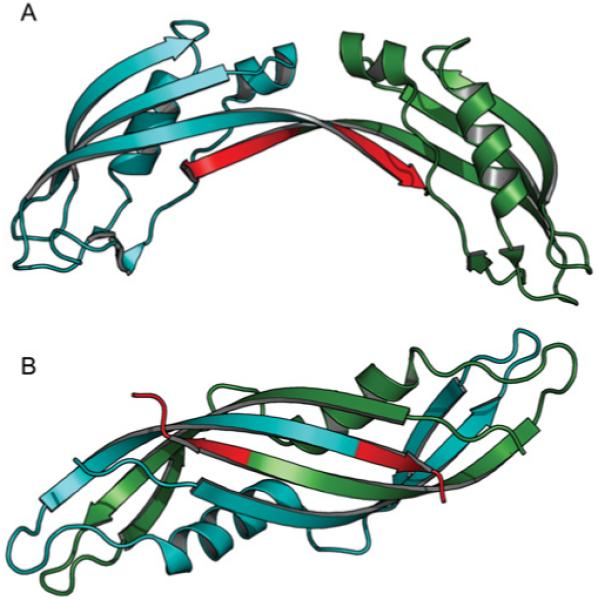
Subunit A is coloured blue and subunit B is coloured dark green. Residues 235–239 are highlighted in red to emphasize which residues are absent in TonB2 from V. anguillarum. (B) Crystal structure of an intertwined dimer form of TonB-CTD from E. coli [11,12]. The colour scheme is the same as in (A).