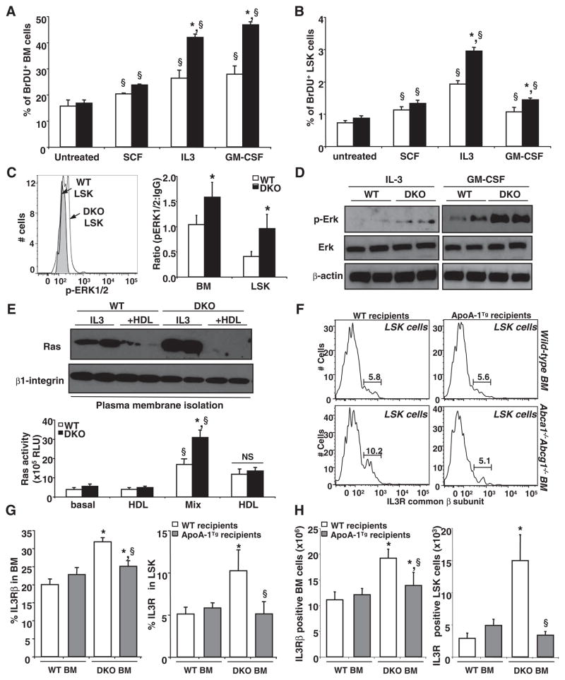
Fig. 3.
HDL protects myeloid cells from activation of IL-3–receptor β canonical pathway. (A) WT and Abca1−/− Abcg1−/− BM cells and (B) LSK cells were grown for 72 hours in liquid culture in the presence of indicated growth factors and were analyzed for BrdU incorporation by flow cytometry. (C) Representative histogram and bar graph showing the expression of phoshpERK1/2 by flow cytometry in freshly isolated BM and LSK cells from WT mice transplanted with control or Abca1−/− Abcg1−/− BM. (D) Western blot analysis shows phospho-ERK, total ERK, and β-actin expression in WT and Abca1−/− Abcg1−/− BM cells treated with indicated growth factors in duplicate samples. (E) Duplicate samples of BM cells treated with indicated growth factors and 50 μg/mL HDL cholesterol were subjected to plasma membrane fractionation and analyzed for Ras and β1-integrin expression or used to quantify Ras activity. (F) Representative histograms showing the expression of the IL-3–receptor β in LSK cells from WT and apoA-I transgenic recipient mice transplanted with control or Abca1−/− Abcg1−/− BM. (G) Percentage and (H) absolute number of BM and LSK cells expressing the IL-3Rβ are depicted from the above-mentioned mice. Results are means ± SEM (error bars) of five to six mice per group. *P < 0.05 genotype effect; §P < 0.05 treatment effect.

