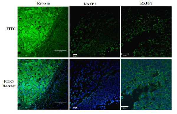
Figure 5.
Validation of antibodies on porcine tissues using immunofluorescence detection. Validations were performed on a minimum of three slides prepared from three independent ovary collections. Sections of porcine ovaries were fixed for immunofluorescence detection of relaxin in corpora lutea and RXFP1 and RXFP2 in follicles. Micrographs indicate the labeling of protein targets (FITC, green color) in the luteal (relaxin) and granulosa and theca cells (RXFP1 and RXFP2). Both receptor signals are mostly located in the region of plasma membrane, while relaxin signal appears homogenous within the cytoplasm. Relaxin staining often delimits a dark hole corresponding to the nucleus stained with Hoechst dye (blue color). Not all cell types showed the FITC signals of protein targets. Scale bars = 100 μm.

