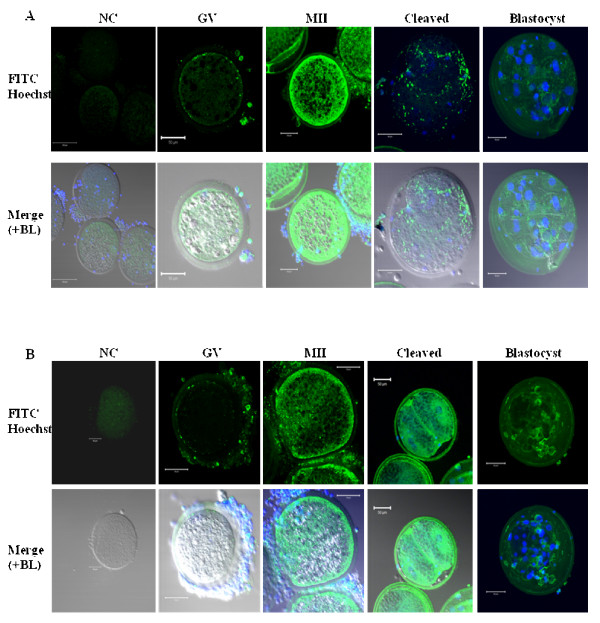
Figure 7.
Immunofluorescence detection of RXFP1 (A) and RXFP2 (B) protein in porcine oocytes and embryos. A minimum of 50 oocytes and embryos derived from 3 independent replicates were analyzed for immunofluorescence (IF). Micrographs show the IF detection (FITC, green color) of RXFP1 (A) and RXFP2 (B). The plasma membrane location of both receptor signals is visible in oocytes, cumulus cells and blastocyst. The FITC signals in tested groups appear above the background as seen in the negative control group (NC). Nuclei are stained in blue with the Hoechst dye. GV = Immature oocytes, MII = metaphase II oocytes, BL = Bright light. Scale bars = 50 μm.

