Figure 2.

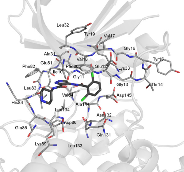
Orientation of the CDK2 active site in the PDB structure 2VU3 showing the amino acid residues (grey lines) used for the QM and MM calculations. Ligand 33 is shown as grey sticks.

Orientation of the CDK2 active site in the PDB structure 2VU3 showing the amino acid residues (grey lines) used for the QM and MM calculations. Ligand 33 is shown as grey sticks.