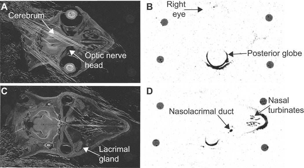
Figure 3. Ocular distribution of 14C-TG100801 following topical instillation in the rabbit.

Transillumination (A & C) and corresponding autoradiograph (B & D) images of cross sections through a rabbit’s head taken 30 minutes after a single unilateral (left eye) topical instillation of 1% TG100801 containing radiotracer levels of 14C-TG100801. A 40 µm section at the horizontal midline (A & B) or inferior to the midline capturing the nasolacrimal duct (C & D). The four circles visible at the perimeter of each tissue section are remnants of mounting posts.
