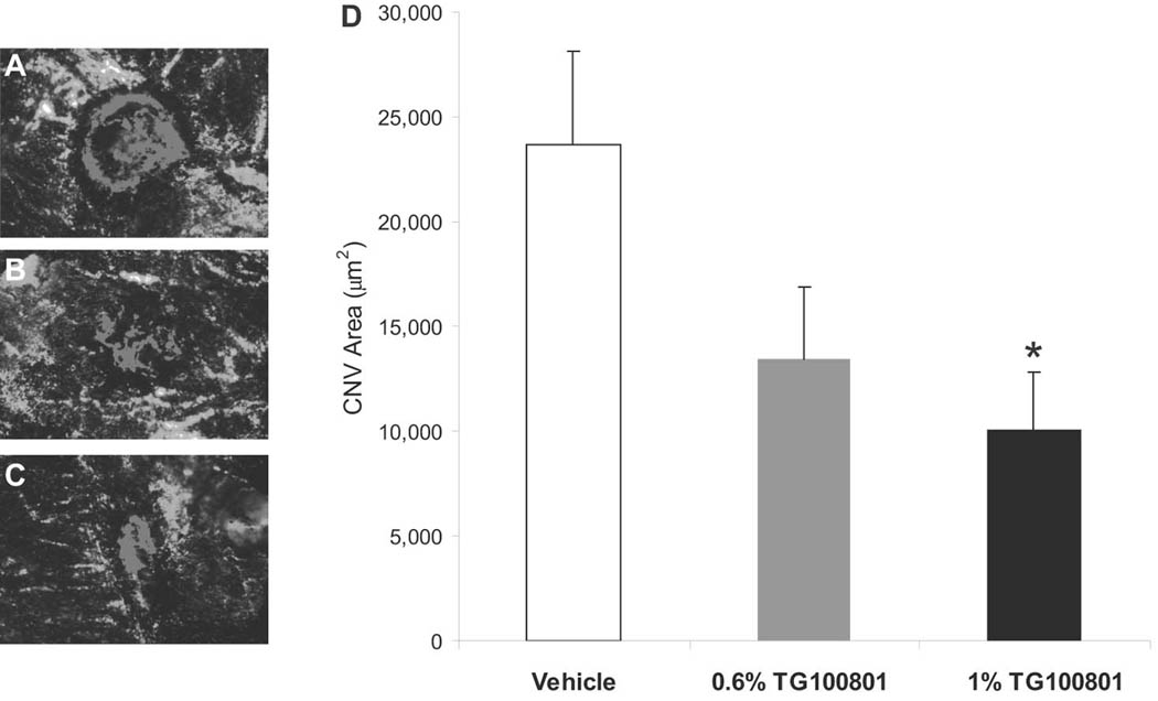
Figure 4. Topical delivery of TG100801 in a murine model of choroidal neovascularlization (CNV).

Laser-induced rupture of Bruch’s membrane was performed in C57BL/6 mice, after which animals were dosed topically twice a day with either TG100801 (1% or 0.6% formulations) or vehicle. After 14 days, animals were perfused with FITC-dextran and choroidal whole mounts were photographed for measurement of CNV area by computerized image analysis. (A–C) Representative fluorescent photomicrographs taken from the three treatment groups (A= vehicle, B= 0.6% TG100801, C= 1% TG100801), with fluorescence of the CNV highlighted in red. (D) CNV area graph (data shown as means ± SEM, n= 24 lesion sites; * 1% TG100801 and vehicle groups differ with P= 0.036).
