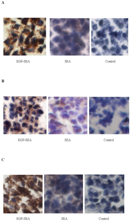
Figure 3. Detection of intratumoral CTLs.
Small brown spots around S180 tumor cells indicated antibody-reactive T cells. (A) CD4-positive cells detected by rabbit CD4-specific polyclonal antibody. (B) CD8 positive cells detected by rabbit CD8-specific polyclonal antibody. (C) SEA-reactive T cells. Sections were incubated with a SEA solution, followed by mouse polyclonal anti-SEA IgG fraction and anti-mouse secondary antibody. Microscope magnification: ×400 in A to C.

