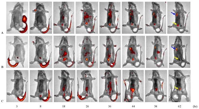
Figure 6. Monitoring of the targeting of EGF-SEA to solid tumors by in vivo imaging of mice.
LSS670-labeled EGF-SEA proteins (125 pmol) were injected i.v. into mice bearing tumors (A and B) and un-inoculated mice as control (C). Movement of labeled EGF-SEA proteins was observed using in vivo fluorescence imaging with the IVIS Kinetic imaging system. Blue and yellow arrows indicated the tumor and bladder positions.

