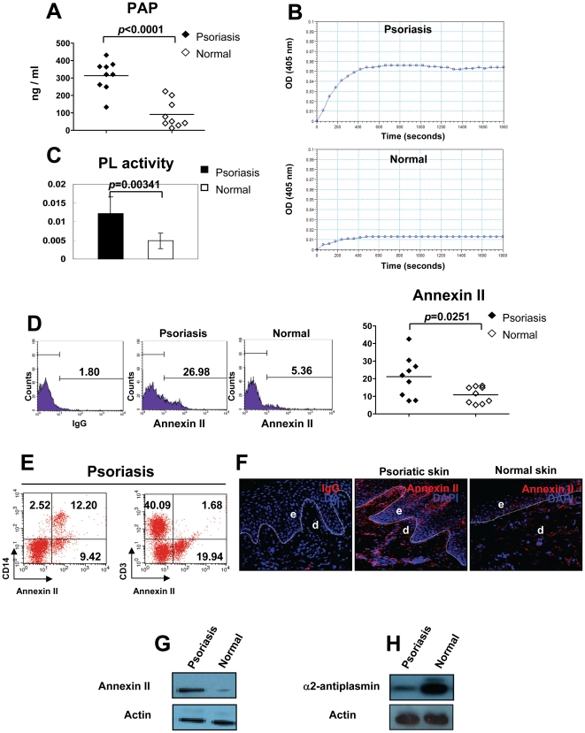
Figure 2. Enhanced PL activity in psoriasis.
(A) PL was measured by ELISA. (B) PL activity was detected by a chromogenic assay. (C) The PL activity was represented by ΔA/min (absorbance/minute). (D) The percentage of annexin II+ cells in PBMC of psoriasis patients and normal controls is shown. (E) Expression levels of annexin II on CD14+ monocytes and CD3+ T cells in PBMC of psoriasis patients was measured by Flow cytometry. One representative experiment out of 5 is shown. (F) Cryosections from lesional skin of psoriasis patients or normal volunteers were stained with an annexin II mAb (red). Original magnification, ×400. e, epidermis; d, dermis. Dotted lines indicate the border between epidermis and dermis. (G, H) Lysates from lesional skin of psoriasis patients or normal skin of volunteers were subjected to western blotting for annexin II expression or α2-antiplasmin expression. Actin - loading control.

