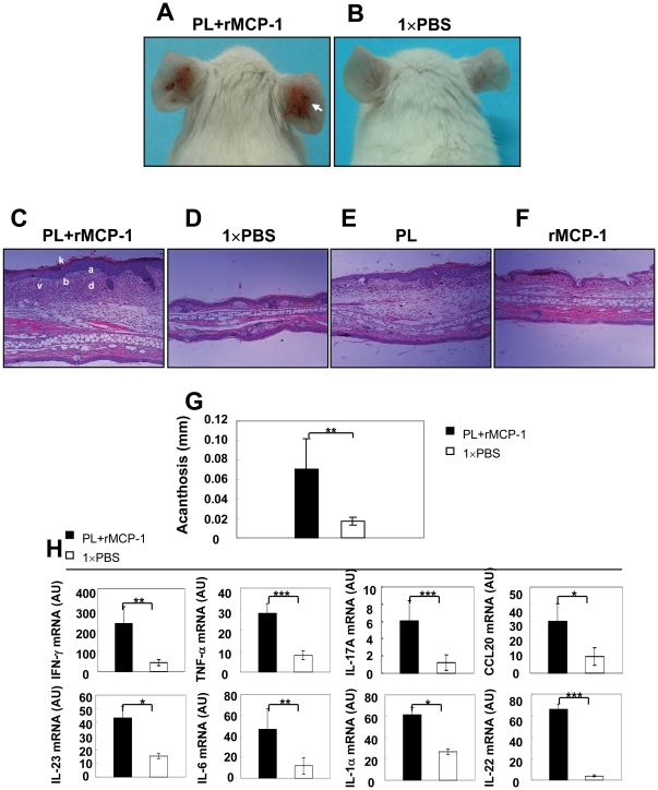
Figure 3. Psoriasiform skin disease induced by PL in mice.
The ears of WT BALB/c mice were injected intradermally every other day for 28 days with 30 µl 1×PBS either containing 1.5 µg PL and 30 ng recombinant murine MCP-1 or alone. (A, B) Representative clinical features of ears of treated mice. White arrow indicates severe erythema, scales and crusts. (C–F) H&E staining of ears of from mice treated with PL+rMCP-1, 1×PBS, PL and rMCP-1. k, hyperparatosis with intracorneal neutrophilic pustule; a, acanthosis; b, increased proliferative basal layer epidermal keratinocytes; d, dermal cell infiltrate; v, dilated dermal blood vessels. Original magnification, ×100. (E) Microscopic changes of ears quantified using acanthosis. (F) Real-time RT-PCR analysis of ears with indicated treatments. * p<0.05, ** p<0.01, *** p<0.001, Student's t test.

