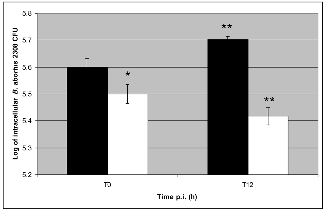
Figure 1. Kinetics of B. abortus S2308 intracellular growth in bovine monocytes-derived macrophages from cattle naturally resistant (R) or susceptible (S) to brucellosis.
Monocyte-derived macrophages (MDMs) were plated in triplicate in 24 well plates at 2 × 10⁵ cells/well and infected with B. abortus S2308 at a MOI 5:1. After 30 min – interaction, extracellular bacteria were killed by co-incubation with gentamycin for 1 h, and then washed 3 times with PBS. At 0 (T0) and 12 (T12) h post-infection, cells were lysed and serial dilutions cultured on TSA plates for quantification of CFU. The intracellular number of B. abortus S2308 was significantly different (P < 0.05) in MDMs from R and S cattle at T0 (*) and at T12 from R or S MDMs compared to their own T0 value (**). Means +/− SD (bars) of 3 independent assays done in triplicate are shown. Solid bars indicate intracellular B. abortus CFU from S MDMs, open bars indicate intracellular B. abortus CFU from R MDMs.

