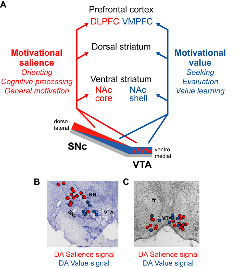
Figure 7. Hypothesized anatomical location and projections of dopamine motivational value and salience coding neurons.
(A) In our hypothesis, motivational salience coding DA neurons are located predominantly in the dorsolateral SNc and medial VTA. They may send signals to regions of the nucleus accumbens core (NAc core), dorsal striatum, and dorsal and lateral prefrontal cortex (DLPFC). Motivational value coding DA neurons are located predominantly in the ventromedial SNc and throughout the VTA. They may send signals to regions of the nucleus accumbens shell (NAc shell), dorsal striatum, and ventromedial prefrontal cortex (VMPFC).
(B) DA excitatory responses to aversive cues (red dots) often occur in the dorsolateral SNc, while inhibitory responses (blue dots) often occur in the ventromedial SNc. Data are from one monkey and collapsed across three adjacent 1 mm sections. Also labeled are the substantia nigra pars reticulata (SNr) and red nucleus (RN). Adapted from (Matsumoto and Hikosaka, 2009b).
(C) DA neurons with greater excitation (red dots) or inhibition (blue dots) to aversive cues than neutral cues are mixed within the medial VTA. Also shown are neurons that had greater responses to neutral cues than aversive cues (gray dots). Also labeled is the fasciculus retroflexus (fr). Data are from eight rabbits and collapsed across three adjacent sections. Adapted from (Guarraci and Kapp, 1999).

