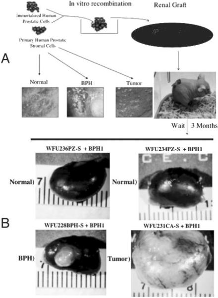
Fig. 5.
BPH-S and CA-S cells induce prostate epithelial growth. A, Schematic of prostate tissue recombination protocol. Isolated prostatic stromal cells of defined histological origin (representative toluidine-stained sections are depicted) are combined with BPH-1 cells in vitro in a collagen gel matrix. The recombinant button is grafted under the renal capsule of a nude mouse and allowed to grow for 3 months. B, Representative photomicrographs of dissected kidneys grafted with prostate tissue recombinants. The stromal strain used for each graft is indicated above the corresponding photomicrograph.

