
Figure 2.
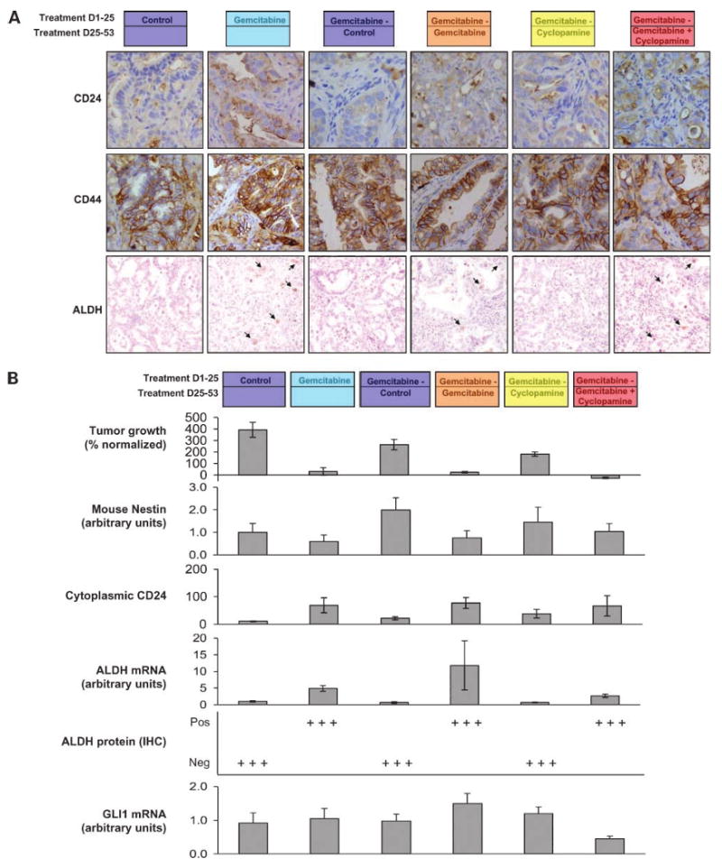
A, immunohistochemical assessment of CD24, CD44, and ALDH by immunohistochemistry. CD44 presented a flat staining that was not different between groups. However, both CD24 and ALDH had a pattern consisting in inversely intense staining with tumor size, with maximal staining in gemcitabine-treated and gemcitabine + cyclopamine-treated groups. B, tumor growth, mouse nestin expression, cytoplasmic CD24, ALDH by reverse transcription-PCR and immunohistochemistry, and GLI1 mRNA expression. ALDH by immunohistochemistry was particularly differentiating as the low-level expression followed an all-or-nothing pattern. ALDH was only positive in 2% of the cells in the gemcitabine-treated groups. In the group where gemcitabine is ceased, the TSC markers (both CD24 and ALDH) decreased back to the levels of the untreated group, suggesting a repopulation of proliferating cells and a dilution of the TSC-enriching effect.