
Figure 2. Immunostainings of kidney sections of a Connexin 40 (Cx40)fl/fl mouse (control) and renin-Cre Cx40fl/fl mouse.
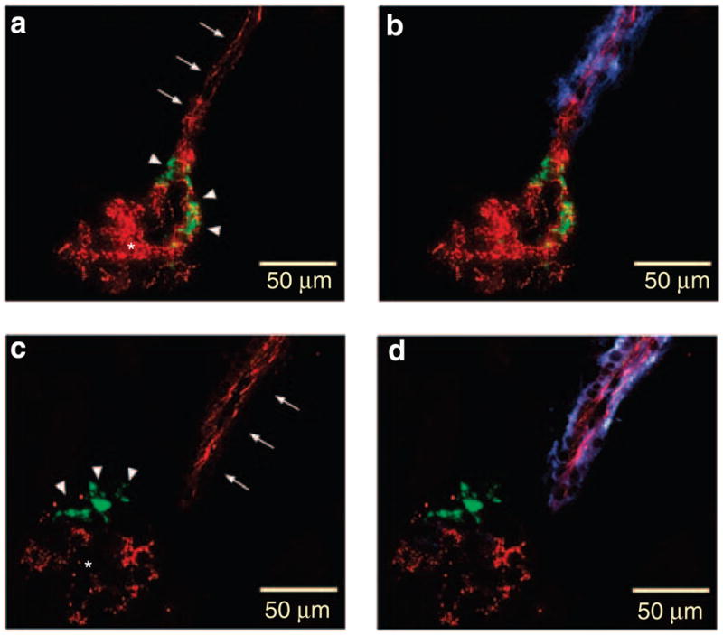
(a, c) are immunostained for Cx40 (red) and renin (green). b and d are immunostained for Cx40 (red), renin (green), and α-smooth muscle actin (blue) as a smooth muscle cell marker in an afferent arteriole. Arrows indicate endothelial staining, arrowheads renin-producing cells and asterisks intraglomerular mesangial cells. Note the coexistence of Cx40 and renin immunoreactivity in the Cx40fl/fl kidney (a, b), whereas Cx40 immunoreactivity is absent in the renin-Cre Cx40fl/fl kidney from renin-producing cells (c, d). Also, note the similar Cx40 immunoreactivity in the endothelium both in Cx40fl/fl (a, b) and in renin-Cre Cx40fl/fl (c, d) kidneys.