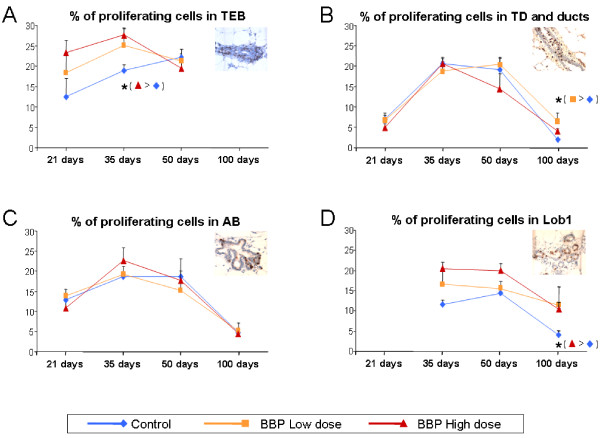
Figure 2.
Proliferative index in the mammary gland. Percentage of proliferating cells in TEB (A), TD and ducts (B), AB (C) and Lob1 (D) at different ages in the mammary gland of rats exposed prenatally to low or high dose of BBP. Proliferating cells were identified by immunohistochemical detection of BrdU incorporation (brown cells). Olympus BX40 microscope with 40x objective. *: significant differences (p < 0.05).

