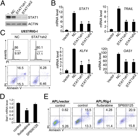
Fig. 5.
RIG-I induction triggers ISG production and growth inhibition of AML cells partly through its promoting role on STAT1 activation. (A) Two shRNA plasmids specific for STAT1 knockdown (STAT1sh1 and STAT1sh2) and one plasmid containing a negative control (NC) sequence were individually transduced into 293T cells, together with STAT1-expressing plasmid. Forty-eight hours later, the cell lysates were collected for STAT1 protein analyses by Western blotting assay. (B and C) Two STAT1 shRNA plasmids and one control plasmid were individually transduced into U937/RIG-I cells at day 2 after tetracycline withdrawal and then cultured in the Tc− medium for another 4 d. (B) Relative mRNA levels of STAT1 and ISGs as indicated were determined by real-time PCR assays (n = 3, mean ± SD; *P < 0.05). (C) Cell cycle measurement by propidium iodide (PI) staining or cell death analysis by Annexin V-PI staining was performed by flow cytometry. (D and E) RIG-I–transduced mouse APL BM cells were treated with 100 μM fludarabine (Stat1 inhibitor) or 5 μM SP600125 (JNK inhibitor) for 48 h. Cells were then measured by real-time PCR for Stat1 mRNA level (n = 3, mean ± SD; *P < 0.05) (D) or analyzed by flow cytometry for cell survival (E).

