Fig. 5.
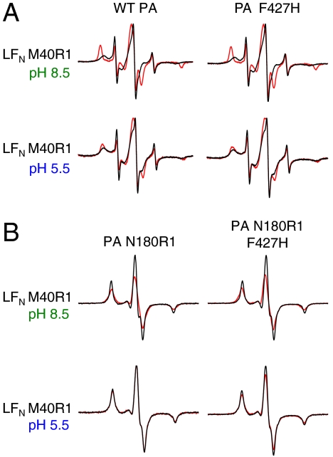
LFN residue 40 interacts with PA α-clamp. (A) EPR spectra of LFN M40R1 with and without WT PA or PA F427H. Black line—LFN M40R1 alone. Red line—with WT PA or PA F427H (1∶1 molar ratio LFN∶PA heptamer). (Top) Spectra collected at pH 8.5 (when PA is in the prepore conformation). (Bottom) Spectra collected at pH 5.5 (when PA is in the pore conformation). Spectra were collected at room temperature and normalized to the intensity of the central peak. (B) Black—additive EPR spectrum of LFN  N180R1 or PA N180R1 F427H. Red—EPR spectrum of 1∶1 molar ratio LFN M40R1:PA N180R1 or LFN M40R1:PA N180R1 F427H complex. Spectra were collected at 233 K either at pH 8.5 (when PA is in the prepore state) or pH 5.5 (when PA is in the pore state) and normalized to the same number of spins.

