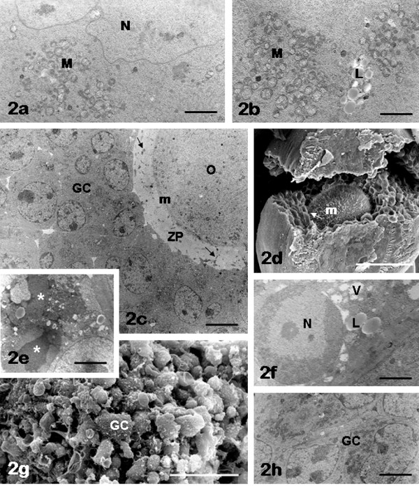
Figure 2.

FCS follicles: oocyte and granulosa cells. Note the presence of juxta-nuclear aggregates of mitochondria (M) and lipid droplets (L) in the oocyte (panels a, b). Scarce (panel c) or more abundant (panel d) microvilli (m) are present on the oolemma. Damaged granulosa cells (asterisks, panel e) are seen around the oocyte, sometimes showing chromatin margination in the nucleus (N), vacuoles (V) and lipid droplets (L) in the cytoplasm (panel f). A diffuse blebbing on the granulosa cell (GC) surface is also observable in panel g. Granulosa cells (GC) with normal appearance are clearly recognizable in panels c, h. Panel a, N: oocyte nucleus; panels c, d, arrows: granulosa cell cytoplasmic projections crossing the zona pellucida (ZP) to reach the oocyte (O) surface (transzonal processes). Bar is: 2 μm (panel a); 1.8 μm (panel b); 5 μm (panel c); 52 μm (panel d); 2.8 μm (panel e); 1.5 μm (panel f); 40 μm (panel g); 2.1 μm (panel h). Panels a-c, e, f, h: TEM; panels d, g: SEM.
