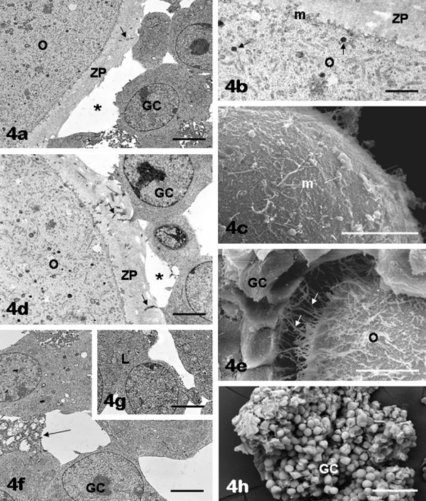
Figure 4.

FSH antral follicles: oocyte and granulosa cells. The oocytes (O) contain numerous organelles, uniformly distributed in the ooplasm (panels a, b, d). Note the presence of scattered cortical granules (arrows) in the suboolemmal area (panel b). Numerous microvilli (m) can be observed on the oocyte surface (panels b, c). Cytoplasmic projections (arrows) stemming from the inner granulosa cell (GC) layer are seen crossing the zona pellucida (ZP) (panels a, d) and reaching the oocyte (transzonal processes) (panels d, e). Areas of detachment between inner granulosa cells and oocyte are also present (asterisks, panels a, d). Outer granulosa cells (GC) appeared irregularly rounded/polygonal and scarcely adherent each other (panels f, h). Uniformly dispersed chromatin and one or more nucleoli are seen in both inner (panels a, d) and outer (panels f, g) granulosa cells. Panel b, ZP: zona pellucida. Panel e, O: oocyte. Panel f, arrow: loss of contact among granulosa cells; panel g, L: lipid droplets in the granulosa cell cytoplasm. Bar is: 3.5 μm (panel a); 1 μm (panel b); 10 μm (panel c); 2 μm (panel d); 9 μm (panel e); 3 μm (panels f, g); 50 μm (panel h). Panels a, b, d, f, g: TEM; panels c, e, h: SEM.
