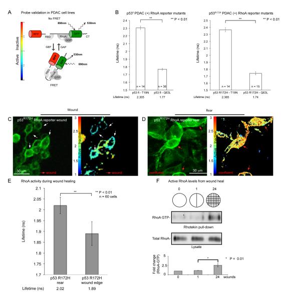
Figure 1.
Fluorescence life-time imaging of RhoA activity within a migrating pancreatic tumor cell population during wound healing. A, Schematic of the adapted GFP-RFP Raichu-RhoA reporter adapted from Yoshizaki et al (4). B, Quantification of life-time measurements in p53fl or p53R172H PDAC cells transiently transfected with dominant negative (T19N) or constitutively active (Q63L) mutants of the Raichu-RhoA reporter. C, D Representative fluorescence image of p53R172H PDAC cells expressing the Raichu-RhoA reporter (green) at the front or rear of a wound with corresponding life-time maps of RhoA activity. White arrows depict active cells. E, Quantification of life-time measurements of RhoA activity for cells at the front or rear of the wound. Cells were classed to be at the front of the wound within the first three cells from the wound border. F, Anti-Rho immunoblot of total cell lysate and GST-Rhotekin ‘pulldown’ (GTP-Rho) from confluent versus single or multiple wounded p53R172H PDAC cells. Columns, mean; bars, SE. **, P < 0.01

