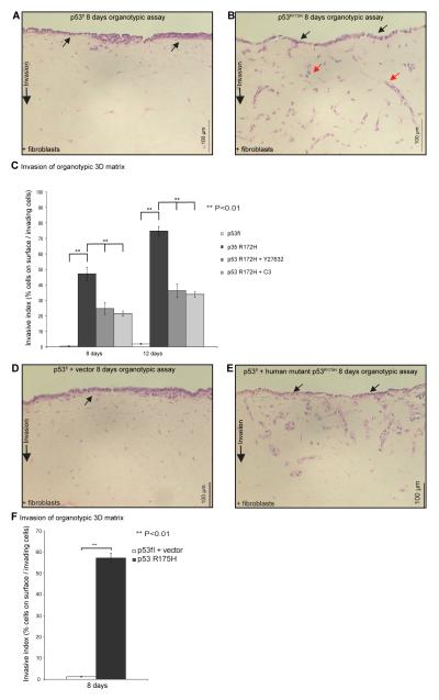
Figure 2.
RhoA activity is required for PDAC invasion. A,B H&E-stained sections of p53fl and p53R172H cells on organotypic matrix. C, Quantification of p53fl and p53R172H PDAC cell invasion ± Y27632 or cell-permeable C3 in the organotypic matrix at 8, 12 days. D,E H&E-stained sections of p53fl cells expressing vector or human mutant p53R175H cultured on organotypic matrix. Quantification of human mutant p53R175H-driven PDAC invasion in the organotypic matrix at 8 days. Red arrows depict examples of cells assessed at depth for Fig. 3C. Columns, mean; bars, SE. **, P < 0.01

