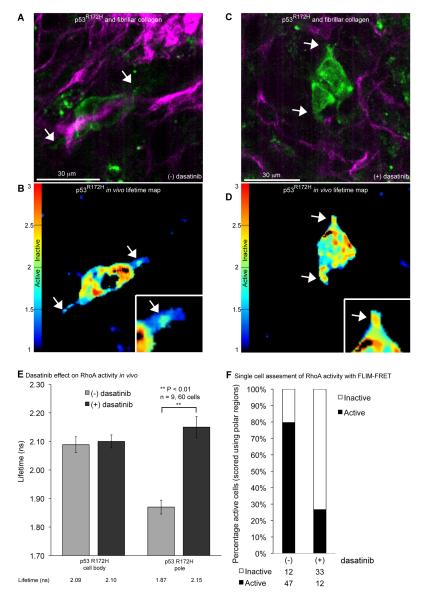
Figure 6.
Spatial regulation of RhoA activity in invasive p53R172H PDAC cells upon dasatinib treatment in vivo. A and C, Representative in vivo fluorescence images of mutant p53R172H PDAC cells expressing the Raichu-RhoA reporter (green) with SHG signal from host ECM components (purple). B and D, Corresponding in vivo life-time maps demonstrating the presence and absence of RhoA activity in sub-cellular polar regions of cells ± dasatinib, respectively. E, Quantification of fluorescence life-time measurements of RhoA activity within the cell body or poles of p53R172H cells ± dasatinib. (F) Distribution of active and inactive polar regions in p53R172H PDAC cells within the tumor tissue ± dasatinib. Columns, mean; bars, SE. **, P < 0.01

