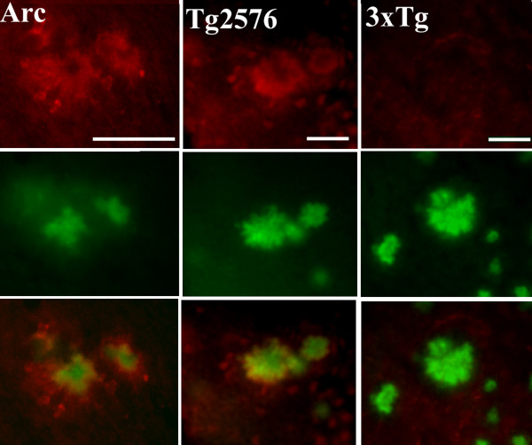
Figure 4.
C1q deposition is not detected on plaques in 3xTg in contrast to other APP mouse models. Representative pictures of C1q (red) and thioflavine (green) and merge (bottom row) show colocalization in Arc48 mice at 9 m (left) and Tg2576 at 18 m (middle), with no detectable staining of C1q in 3xTg at 22 m (right). Scale bar: 50 um.

