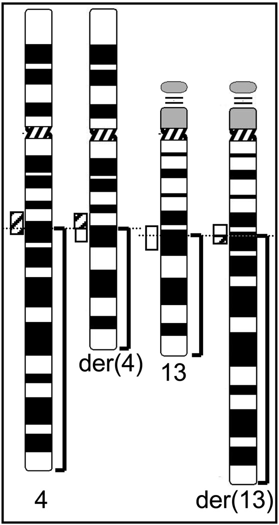
Fig. 2.
A) Schematic diagram of the karyotypic abnormalities in case T-0512 initially reported as t(4;13)(q21.3;q21.2). Dotted horizontal lines indicate the approximate breakpoint locations at 4q22.1 and 13q21.3 as determined by FISH, and translocated parts are bracketed. The hatched and open boxes represent the breakpoint-spanning probe contigs for chromosome 4 and 13, respectively. Please note that the contigs are split unevenly by the translocation.

