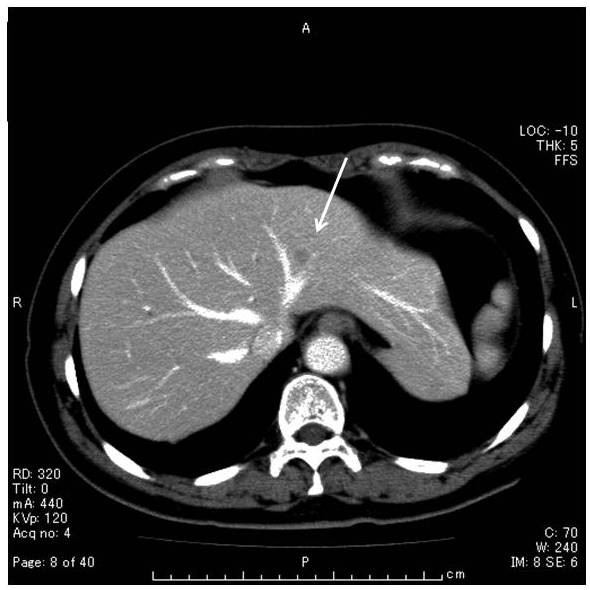
Figure 1.

Unenhanced CT scan showed low density area of 1 cm in diameter in the segment 3 of the liver (arrow). Contrast-enhanced CT scan during arterial phase showed minimally peripheral ring enhancement. No lymphadenopathy or hepatosplenomegaly was observed.
