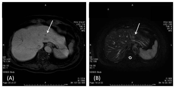
Figure 2.

Magnetic resonance imaging (MRI), the hepatic tumor (arrow) was low signal intensity in T1-weighted image (A) and slight high signal intensity in T2-weighted image (B), and low signal intensity in hepatobiliary phase after Gd-EOB-DTPA injection, and on dynamic Gd-EOB-DTPA MRI protocol not clearly visualized during arterial dominant phase with slight ring-like enhancement persisting, indicating hypovascular tumor, such as cholangiocarcinoma or liver metastasis.
