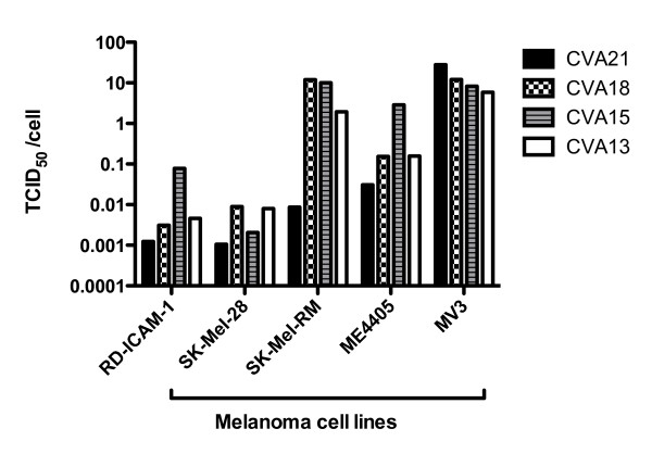
Figure 2.
Destruction of melanoma cells by CVA13, CVA15, CVA18 and CVA21. Cultures of SK-Mel-28, SK-Mel-RM, ME4405, MV3 and RD-ICAM-1 monolayers were infected with 10-fold serial dilutions of virus ranging from 1:10 to 1:107. After incubation for 48 h, the plates were fixed and stained with a crystal violet/methanol and TCID50/cell required to induce monolayer destruction was calculated. SK-Mel-28 and RD-ICAM-1 were the most sensitive lines to the panel of CVAs requiring only low concentrations of virus to achieve cell lysis.

