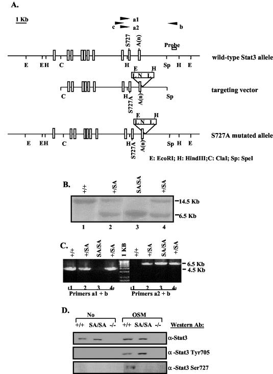
FIG. 1.
Generation of the Stat3 S727A mutant mice. (A) Schematic diagrams of the mouse endogenous wild-type Stat3 allele, S727A targeting vector, and the resulting S727A mutant allele after homologous recombination. Exons are shown as rectangles. Primers a1 and a2 were matched to wild-type serine 727 or mutated alanine sequences, respectively, and were used in pairs with primer b to perform PCR screening for mutation. Primers b and c were used to PCR amplify the fragment in between, which was sequenced to confirm the S727A mutation. N, Neo cassette; A(n), polyadenylation sequence; L, loxP sequence. (B) Southern blot analysis of EcoRI-digested genomic DNA from +/+, SA/+, and SA/SA mice using the probe shown in panel A. The probe detects a 14.5-kb band from the wild-type allele and a 6.5-kb band from the mutant S727A allele. (C) PCR analysis of the genomic DNA analyzed in panel B. Primers a1 and b detect the ∼4.5-kb band from the +/+ allele, and primers a2 and b detect the ∼6.5-kb band from the mutant S727A allele. (D) Western blot analysis to check serine phosphorylation. Whole-cell extracts prepared from +/+, SA/SA, or −/− MEFs untreated or treated with OSM for 30 min were immunoprecipitated with an anti-STAT3 antibody, followed by Western blotting using an antibody against phospho-STAT3 (Ser727). The same extracts were also straightly probed with an anti-phospho-STAT3 (Tyr705) antibody or stripped and reprobed with an anti-STAT3 antibody.

