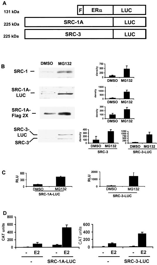
FIG.1.
SRC-1A-LUC and SRC-3-LUC fusion proteins behave as their unmodified counterparts do. (A) Schematic of luciferase fusion constructs. The luciferase (LUC) cDNA was fused to the C terminus of FLAG (F)-ERα, SRC-1A, or SRC-3. The predicted molecular masses are listed on the left. (B) Western analysis of endogenous (SRC-1) and transfected (1,000 ng of SRC-1A FLAG 2× or 1,000 ng of SRC-1A-LUC) SRC-1 in HeLa cells in the absence (DMSO) or presence of 10 μM MG132 for 24 h was performed using appropriate antibodies (see Materials and Methods). The histograms to the right represent densitometric quantitation of Western blots from two or three separate experiments. (C) The luciferase activity of 100 ng of SRC-1A-LUC or 250 ng of SRC-3-LUC transfected into HeLa cells and treated with MG132 or DMSO vehicle for 24 h. (D) SRC-1A-LUC and SRC-3-LUC can coactivate ERα. HeLa cells were transfected with 500 ng of pERE-E1b-CAT, an expression vector for wild-type ERα (10 ng of pCR3.1 hERα) along with 250 ng of the pCR3.1 empty vector (−), pCR3.1 SRC-1A-LUC, or pCR3.1 SRC-3-LUC and treated with either ethanol vehicle (−) or E2 for 24 h and assayed for CAT protein levels.

